Using 3D Reconstructions of Organs for Pre-Surgical Planning
by Madhu Reddiboina | Aug 30, 2021 | Health Care

Using 3D Reconstructions of Organs for Pre-Surgical Planning
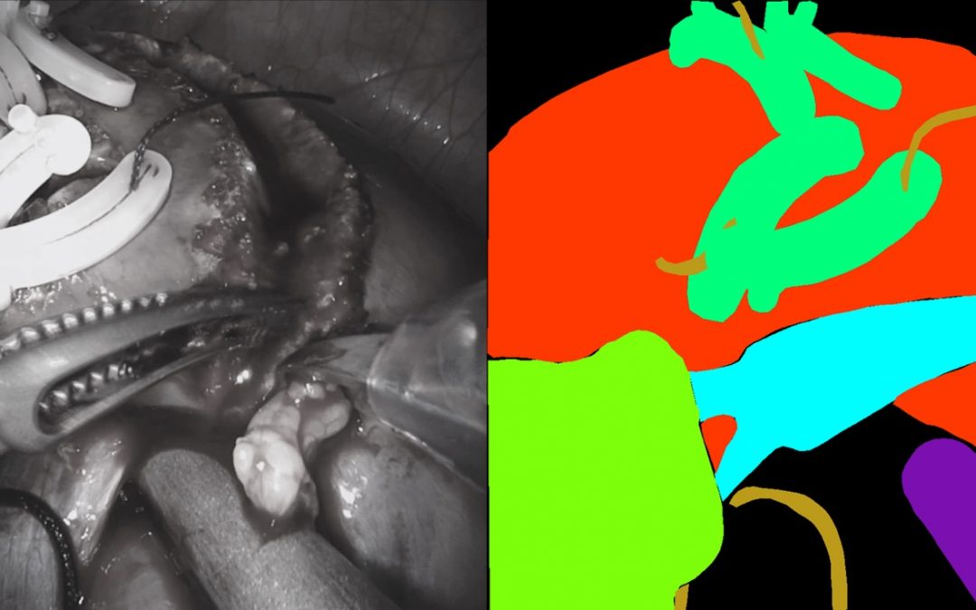
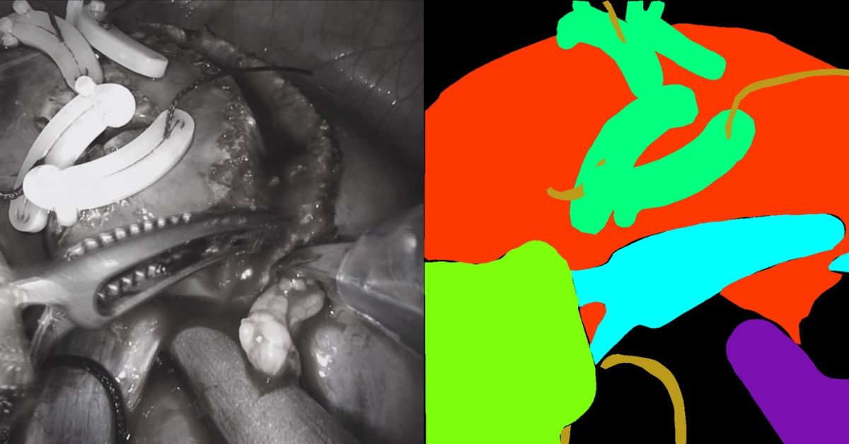
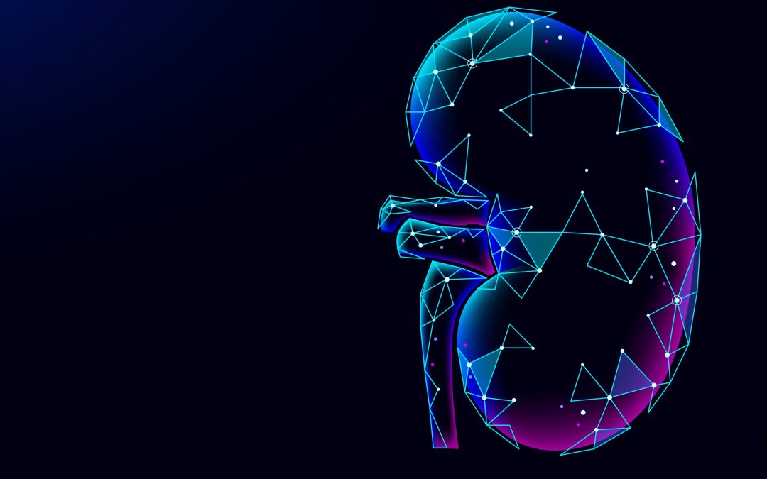
RediMinds took on the challenge of creating 3D models of anatomical objects. Using deep learning algorithms, the RediMinds team developed an automated model for semantic segmentation of a kidney and tumor. By filtering out the surrounding tissue and identifying the tumor areas, the platform was able to construct a 3D model of the kidney with the tumor, allowing the model to be viewed from a 3D perspective.
How 3D Reconstruction of Organs is Beneficial
As stated earlier, 2D images, such as CT scans and x-rays, have primarily been used for pre-surgical planning in procedures dealing with soft tissue-based procedures. However, with 3D technology, surgeons can get a better idea of the anatomical structure of the organ and the location of the organ relative to other soft tissue. With this invaluable insight, surgeons can create a route for the surgery and plan the procedure with more efficiency.
When only relying on x-rays or CT scans, there is a potential for valuable information to be missed during the pre-operative planning process. This could potentially slow down or create issues during the procedure. When using a 3D model of an object, the surgeon can easily rotate and view the object from many different angles. If printed, the object can be held and observed from a realistic perspective, which can give insight into the size of the object. Pre-operative planning using 3D models can lead to better diagnostic accuracy.
Furthermore, this technology enables more collaboration between surgeons. With 3D models, it is possible for teamwork to happen regardless of geography. For example, a surgeon who lives in another country can easily access a 3D model to diagnose a problem and plan an operation. This creates the possibility of experts in certain topics becoming more accessible when their advice would be beneficial for diagnosing or treating a patient.
AR/VR Surgical Solutions
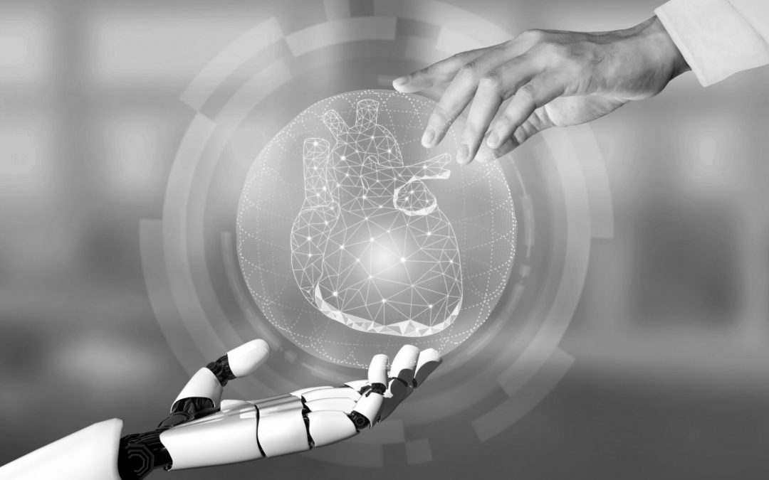
Aside from 3D printing, 3D models could be made for the liver, lungs, or heart. These 3D models could provide the basis for AR/VR reconstruction of the anatomical object. Anatomical knowledge of the operative site is essential for soft tissue-based surgeries. VR and AR offer new ways to view three-dimensional anatomical structures for pre-operative planning.
VR allows the surgeon to evaluate and get familiar with complexity of the surgical area and object in a virtual environment. On the other hand, AR allows 3D objects to be projected onto the patient both before and during surgery. This technology could provide insight that could help the surgeon operate faster with more accuracy than when planning operations using only 3D images.
The Future of 3D Reconstruction of Anatomical Objects
Pre-surgical planning is critical to achieving the best possible surgical result and minimizing any problems during or after the surgery. While there are limits to planning with 2D images, creating 3D models of anatomical objects could result in more efficient pre-operative planning and, therefore, better outcomes for patients. This technology provides the building blocks towards a broader solution of developing AI-assisted surgical tools to provide better overall surgical outcomes.
To learn more, check out the case study.

